
Current estimates place the number of migraine sufferers in the United States at approximately 40 million! Sadly, this number does not take into account those who suffer from other types of headaches such as cluster headaches and tension headaches. Fortunately, there is now a well-established, outpatient surgical procedure that has been shown in numerous studies to significantly reduce the frequency, severity and/or duration of migraine headaches and in some cases, to eliminate them permanently.
MIGRAINE/HEADACHE SURGERY

IS PERMANENT RELIEF FROM CHRONIC HEADACHES POSSIBLE?”
YES!
Migraine headaches have traditionally been thought to begin within the central nervous system (i.e., the brain and/or spinal cord) and then produce symptoms elsewhere such as throbbing in the back of the head, forehead, or temples. There are many theories as to what exactly within the central nervous system is causing these chronic and often debilitating headaches. Some of these theories include pathologic blood vessel dilatation and constriction (loosening and tightening), abnormal firing of neurons within the brain, and abnormalities of various biologic substances (e.g. serotonin, calcitonin gene-related peptide). The fact that no one theory has been proven correct is likely one of the many reasons that there are so many different methods for the treatment of chronic headaches like migraines. In fact, from a medication standpoint alone, there are not only dozens of medications used to treat migraines, but dozens of classes of medications such as triptans, anti-depressants, muscle relaxants, blood pressure medications, narcotics, neuroleptics, ergotamines, and so on. About 24 years ago, a different perspective on chronic headaches was re-established and has produced remarkably good results.
This different school of thought suggests that peripheral nerve irritation (i.e., irritation of nerves outside of the brain and spinal cord such as those within the scalp or forehead) can cause irritation within the central nervous system thus leading to the perception of and symptoms of a headache. If this mechanism were in fact the culprit, then identifying and correcting the cause of such irritation could produce relief from the headache symptoms. Plastic surgeons have been doing exactly that with a common nerve irritation problem known as carpal tunnel syndrome for decades. In this syndrome, a nerve within the wrist is compressed (i.e., pinched) and surgeons decompress (i.e., un-pinch) it thereby relieving the symptoms of pain with a greater than 90% success rate. Numerous research studies have definitely demonstrated that just like at the wrist, there are nerves within the head and neck that are compressed and that decompressing them, can produce significant or even complete relief that can be permanent.
HOW DO I KNOW IF A PINCHED NERVE IS THE CAUSE OF MY HEADACHES?”
During your consultation with Dr. Peled, he will take a thorough history focusing specifically on your headache symptoms. We will discuss all the treatments which you have tried in the past and go over any old imaging studies. Dr. Peled will then perform a comprehensive physical examination to elucidate if there are any physical findings that suggest that a pinched nerve in the head or neck could be causing your headache. Additional imaging studies (i.e., MRIs or CT scans) may be required and if so, they will be ordered accordingly. If appropriate, a series of diagnostic nerve blocks – simple injections with local anesthetic (i.e., a numbing agent) – will then be performed in the office. Depending on the results with these various steps, a decision will be made as to whether your headaches could be caused by a pinched nerve and hence your suitability for surgical decompression.
WHY USE NERVE BLOCKS?
Simply put, a nerve block chemically and temporarily inactivates a nerve. Anyone that has been to the dentist for a cavity knows what a nerve block is – the numbing agent used to numb the tooth so that a filling can be performed. If a sensory nerve (one that provides sensation to a part of the body) is irritated, it can cause pain. When that nerve is inactivated, the pain improves. Therefore, if you have a severe headache caused by an irritated nerve, blocking that nerve will temporarily relieve that headache. It is important to remember that nerve blocks are diagnostic, not therapeutic. They are like the MRI you get if you hurt your knee skiing. The MRI may show that you’ve torn your ACL, but it will not fix the ACL – that operation needs to be done by an orthopedic surgeon. In the same way, the relief of the blocks will be temporary and hence will not fix the problem. However, they are excellent tools to help figure out which nerves are causing the headache pain and should be addressed surgically. These diagnostic injections must be performed in a very specific way, by someone with a thorough understanding of the anatomy which is being addressed. Having operated on thousands of nerves in the head and neck, specifically the ones often targeted with these blocks, Dr. Peled has an intimate understanding of the anatomy of the peripheral nerves within the head and neck and has performed numerous successful blocks.
IF I’M A CANDIDATE FOR THIS TYPE OF SURGERY, HOW IS IT PERFORMED?
Surgical decompression for chronic headaches is performed as an outpatient procedure at an accredited surgery center or in the outpatient department of the California Pacific Medical Center. The procedures can last anywhere from 1 hour to 3 hours depending on the number and location of the nerves being treated. Small incisions are made in the area of the nerves and the nerves are identified. The structures causing the compression are removed and, in some cases, if the nerves are very injured, the nerves are cut and placed in the nearby muscles to shut them down. Because these are sensory nerves, no loss of function occurs, and numbness is often temporary as the remaining nerves grow into the initially numb areas. There are few restrictions following these procedures and discomfort is usually very well tolerated with oral pain medication.
WHAT ARE THE RESULTS WITH THIS TYPE OF SURGERY?
As noted above, the results with these types of procedures have been quite impressive. In one study out of Georgetown University, data from 190 patients with pain/headaches in the back of the head who underwent surgical decompression were analyzed. Over 80% of patients experienced at least 50% pain relief and over 43% of patients experienced complete relief of their headaches! In February 2011, the five-year results of such procedures were published in the medical journal, Plastic and Reconstructive Surgery. These results demonstrated that five years following their operation, 88% of patients were still reporting greater than 50% improvement in their headache symptoms and 29% were completely headache-free! There are now over 100 peer-reviewed articles in multiple medical journals (a number of them authored by Dr. Peled)(1-9) that demonstrate similar results and the American Society of Plastic Surgeons even has a formal position statement stating that these operations are not only safe and effective, but should be considered standard of care when conventional treatment modalities have failed.
Dr. Peled’s work in this field has culminated in his being elected President of the Migraine Surgery Society, the preeminent group of headache surgeons in the world. He has lectured and taught other surgeons at numerous national and international meetings and operated in several countries in Europe and South America. Ziv is committed to raising awareness and educating others about the life-changing benefits of headache/migraine surgery. While offering exceptional and compassionate care, Dr. Peled’s mission is to bring positive and substantive change to his patients’ lives.
- Peled, Z. M. A Novel Surgical Approach to Chronic Temporal Headaches. Plastic and reconstructive surgery 2016;137:1597-1600. https://journals.lww.com/plasreconsurg/fulltext/2016/05000/a_novel_surgical_approach_to_chronic_temporal.37.aspx
- Peled, Z. M., Gfrerer, L. Introduction to VSI: Migraine surgery in JPRAS open. JPRAS Open 2024;39:217-222. https://www.jprasopen.com/article/S2352-5878(23)00105-5/fulltext
- Peled, Z. M., Gfrerer, L., Hagan, R., et al. Anatomic Anomalies of the Nerves Treated during Headache Surgery. Plastic and reconstructive surgery Global open 2023;11:e5439. https://journals.lww.com/prsgo/fulltext/2023/11000/anatomic_anomalies_of_the_nerves_treated_during.57.aspx
- Peled, Z. M., Pietramaggiori, G., Scherer, S. Anatomic and Compression Topography of the Lesser Occipital Nerve. Plastic and reconstructive surgery Global open 2016;4:e639. https://journals.lww.com/prsgo/fulltext/2016/03000/anatomic_and_compression_topography_of_the_lesser.7.aspx
- Gualdi, A., Cambiaso-Daniel, J., Gatti, J., et al. Selective denervation of the corrugator supercilii muscle for the treatment of idiopatic trigeminal neuralgia purely paroxysmal distributed in the supraorbital and suprathrochlear dermatomes. The journal of headache and pain 2021;22:9. https://thejournalofheadacheandpain.biomedcentral.com/articles/10.1186/s10194-021-01218-6
- Pietramaggiori, G., Scherer, S. S., Peled, Z. M., Wassim, R. Supraorbital Neuroma: A Rare and Unreported Complication Following Blepharoplasty. Journal of reconstructive microsurgery 2015;31:614-616. https://www.thieme-connect.de/products/ejournals/abstract/10.1055/s-0035-1555012
- Saad, M., Connor, L., Hazewinkel, M. H. J., et al. Pearls for Starting a Headache Surgery Practice in Academic and Private Practice. JPRAS Open 2024;39:127-131. https://www.jprasopen.com/article/S2352-5878(23)00100-6/fulltext
- Scherer, S. S., Schiraldi, L., Sapino, G., et al. The Greater Occipital Nerve and Obliquus Capitis Inferior Muscle: Anatomical Interactions and Implications for Occipital Pain Syndromes. Plastic and reconstructive surgery 2019;144:730-736. https://journals.lww.com/plasreconsurg/fulltext/2019/09000/the_greater_occipital_nerve_and_obliquus_capitis.44.aspx
- Amirlak, B., Lu, K. B., Erickson, C. R., et al. In-Depth Look at the Anatomical Relationship of the Lesser Occipital Nerve, Great Auricular Nerve, and Spinal Accessory Nerve and Their Implication in Safety of Operations in the Posterior Triangle of the Neck. Plastic and reconstructive surgery 2020;146:509-514. https://journals.lww.com/plasreconsurg/fulltext/2020/09000/in_depth_look_at_the_anatomical_relationship_of.9.aspx
TREATMENT ZONES

OCCIPITAL

FRONTAL

TEMPORAL
Ziv M. Peled, MD is a Plastic & Peripheral Nerve Surgeon specializing in chronic headache/migraine relief. Board-certified and Harvard-Trained, he is committed to raising awareness and educating others about the life-changing benefits of headache/migraine surgery. While offering exceptional and compassionate care, Dr. Peled’s mission is to bring positive and substantive change to his patients’ lives.
OCCIPITAL
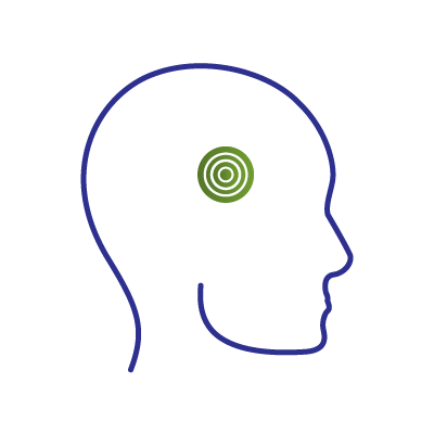
The occipital region is the area in the back of the scalp/head. It is one of the most commonly treated areas for patients seeking surgical relief from chronic headaches. In my humble opinion, many people are often told they suffer from migraines, when they really have neuralgia (i.e. pain caused by mechanical compression or irritation of a nerve). The offending structure(s) causing this compression/irritation may be a spastic muscle, an enlarged or aberrant blood vessel, and/or tight connective tissue. When the pain emanates from and/or occurs in the occipital region of the head, it is called ‘occipital neuralgia’ or ON for short. In the case of people who have pain in the back of the head, the question for the surgeon is, which “neur” (i.e. nerve) is causing the “algia” (i.e. pain).
FRONTAL
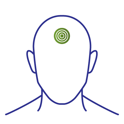
The frontal region is located, as the name suggests, in the front of the head, specifically the forehead and glabella (that region between your eyebrows). When this type of headache pain occurs secondary to nerve compression and/or irritation, it is often secondary to pathology within the supraorbital and/or supratrochlear nerves which are branches of the trigeminal nerve. In some cases, an anterior branch of the zygomaticotemporal nerve (see temporal link) may travel toward the side of the forehead and contribute to the overall, frontal headache complex. It used to be thought that the supraorbital and supratrochlear nerves were primarily compressed by the glabellar musculature, specifically the corrugator supercilii, procerus, and depressor supercilii muscles. However, more recently it has been demonstrated that compression of both nerves may also occur at the orbital rim (i.e. the top of the eye socket) as these nerves sometimes emerge from behind and above the eye through very tight fascial openings or through bony foramina as opposed to notches within the orbital rim. In these locations, adjacent vessels can also act as compressive structures.
TEMPORAL
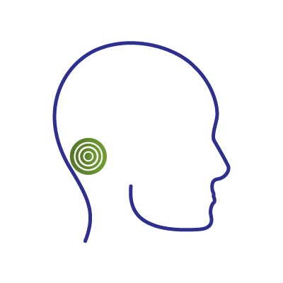
The temporal region is located at the side of the head/scalp – i.e. the temples. Most commonly, headache symptoms in these areas secondary to peripheral nerve irritation/compression are a result of 2 possible, offending nerves. One is called the auriculotemporal nerve and the other is called the zygomaticotemporal nerve and both are branches of the trigeminal nerve. The former is located just above the helical root which is where the external ear (i.e. pinna) meets the scalp and often sends branches ascending toward the vertex of the skull as well as in front of and occasionally behind the ear. In this area, the auriculotemporal nerve may be compressed by the adjacent connective tissue and superficial temporal artery and/or vein which may be enlarged, in an aberrant location, or even wrapping around the nerve itself like an anaconda. The zygomaticotemporal nerve is located just to the side of the eye socket toward the front of the temple. If you clench your back teeth, you might feel a bulge in this area as the temporalis muscle contracts. In this location, the zygomaticotemporal nerve may be entrapped by connective tissue overlying the temporalis muscle which is too tight and/or an adjacent blood vessel. In addition, the zygomaticotemporal nerve may have “downstream” branches that, like the auriculotemporal nerve, go towards the top of the scalp, anteriorly towards the forehead or posteriorly toward the ear. There can be an overlap in the sensory distribution of these two nerves in the mid-portion of the temple which can make it difficult to discern which one of these nerves or both nerves are involved in temporal pain generation.
SCHEDULE A CONSULTATION
We provide a range of emergency, medical, general, residential, and community health services.